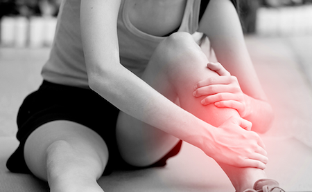
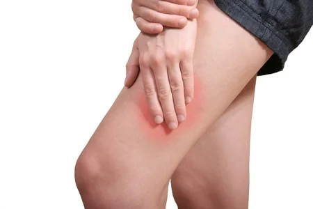
Arthritic Knee

The One Thing That Could Save Your Arthritic Knee

person-holding-knee-in-pain.jpg

In 2014-2015 there were 52,039 knee replacement procedures undertaken in Australia. That represents 257 procedures per 100,000 people aged 18 years and over.

Clearly, this represents an extremely small proportion of people suffering debilitating osteo-arthritic knee pain, most of whom never make it to surgery, yet the reality of living with an osteo-arthritic knee can have significantly debilitating effects on quality of life and income earning capacity [including sport].

THE CAUSES OF OSTEO-ARTHRITIS OF THE KNEE CAN BE ATTRIBUTED TO A MULTITUDE OF FACTORS.

  • Genetic biomechanical anomalies such as knocked knee [genu valgum] or bow legged [genu varum] deformity creates a bias in the weight bearing distribution of the knee joint where only a small portion of the articular surface of the joint is subjected to most of the weight – bearing forces.

  • Repetitive loading will eventually lead to gradual deterioration of the protective articular cartilage surface until it pierces into the underlying pain sensitive bone.

  • Other causes of osteoarthritis often include poorly managed meniscal [cartilage] tears which can occur in a previously healthy knee as the result of a twisting injury in sports or activities requiring directional change such as netball or football or repetitive thrusting with rotation such as occurs in breast stroke swimming.

Understandable confusion exists on the use of the term “cartilage” which may refer to two distinctly different knee structures. The medical term “cartilage” is used to describe the smooth covering on any joint surface which serves to protect bone fr…

Understandable confusion exists on the use of the term “cartilage” which may refer to two distinctly different knee structures. The medical term “cartilage” is used to describe the smooth covering on any joint surface which serves to protect bone from erosion. Colloquially, it is commonly [and incorrectly] used to refer to the meniscus, the crescent shaped pad[s] located between the thigh and shin bones that offer further joint protection.

Tears to a healthy meniscus can occur creating a sharp edge or a small sliver that may not lie entirely congruent with the remaining meniscal body thereby creating a small “step”.

A meniscal fragment can act to both impede locking of the knee as well as shatter the smooth porcelain-like surface of the joint under excessive loading.

For the most part, peripheral meniscal tears generally heal within a couple of months if the split is not being constantly pulled apart by twisting or pivoting on a bent knee.

Significant knee trauma and osteoporosis [de-mineralisation of bones] are also primary contributors to knee osteo-arthritis.

Irrespective of whatever the cause of your osteo-arthritic knee, a common feature is that miniscule cartilage fragments or debris may emerge within the joint obstructing the knee from locking out fully. The greater the joint obstruction, the greater the knee bend and the smaller the contact surface between the femur [thigh bone] and tibia [shin bone].

Continuing to weight bear under these circumstances can precipitate very rapid destruction of a healthy knee leading to pain, swelling and difficulty activating the quadriceps muscles.

The single most important objective should always be to restore full and unrestricted knee extension. Under no circumstances should the knee be forced into a locked or fully straightened position as this will accelerate joint destruction.

TREATMENT:

The most useful technique for restoring knee function to an osteo-arthritic knee is to gently glide the two bones apart with the joint in 90 degrees which can be achieved by placing a webbing belt or folded towel behind the knee and pulling back on the shin bone for a minute or so.

This painless technique can immediately restore knee extension without harming the joint surface.

*INDIVIDUAL RESULTS MAY VARY BETWEEN PATIENTS.

*INDIVIDUAL RESULTS MAY VARY BETWEEN PATIENTS.

Swelling can then be managed through conservative measures such as electrotherapy and anti- inflammatory medication and quadriceps function restored through graduated weight-bearing exercise.

Arthritis

The word “arthritis” comes from the Greek word “arthros” meaning joint. The suffix “itis” refers to inflammation hence “arthritis” simply means “inflammation of a joint”.

The reasons for the joint inflammation are many and varied with close to 100 different types of arthritis identified. Some are associated with systemic (whole body) disease processes such as rheumatoid or other auto immune conditions processes which destroy joint surfaces resulting in chronic pain, stiffness and ultimately impaired function.

The most prevalent type of arthritis is osteoarthritis (OA) in which part or all of a joint erodes over a period of years.

Excessive or asymmetrical weight bearing through a joint over a prolonged period will eventually degrade the smooth articular cartilage covering the joint surface and lead to eventual fragmentation. (Diminished thickness of the protective cartilage eventually exposes unprotected bone).

There are many and varied reasons why some people are more predisposed to OA changes or, in an otherwise healthy individual, some people experience severe OA changes in a solitary joint.

Multiple joint and spinal OA changes may be the result of poor bone density (osteoporosis or osteopoenia) in which the bones are easily degraded. Biomechanical issues such as leg length discrepancy, flat feet, bow legged or knocked knee deformity are prime causative factors for individual joint OA changes.

In isolated cases, some people present with a thickened neck of femur (the connecting bony strut of the “ball” of the hip) which almost always results in hip joint OA early in life. This is known as a CAM deformity and can be identified in clinical examination and confirmed via x-ray.

The most important aspects of successfully managing any of the multiple forms of arthritis are:

·      Early identification of possible biomechanical causes (flat feet, bow legged, knock kneed and leg length
discrepancies) and appropriate correction where possible.

·      Obtaining a correct diagnosis of the type of arthritis and degree of severity of involved joints, to tailor an appropriate
response.

·      Seek appropriate medical and allied health support to prolong normal function.

·      Good nutrition and exercise to optimise bone density, increase joint range and restore muscle supportive strength.

·      Appropriate bracing or orthotic devices to unload affected joints may be useful.

The ageing process inevitably leads to some joint degenerative changes. Maintain a good BMI (body mass index), undertake flexibility and strength work throughout life and seek appropriate professional support to successfully manage arthritis.

Adhesive Capsulitis (Frozen Shoulder)

What is it?

Frozen shoulder is a fairly common condition in the general population. It typically presents in people in their 50s and 60s. Women are more often affected than men.

Basically, it means that your shoulder pain and stiffness is a result of shoulder capsule inflammation and fibrotic adhesions that limit your shoulder movement.

The limitation in movement affects active and passive shoulder joint range of motion. That means that the movement in your shoulder joint is restricted both when you try to move your own arm and when someone else tries to move your arm for you.

Adhesivecap.jpg

What causes it?

Still largely unknown.

Virtually any shoulder condition which causes pain and therefore disuse of the shoulder can increase the likelihood of developing a frozen shoulder.

A potentially preventable cause of frozen shoulder can occur post surgery. Patients who are overly protective of their arm and do not comply with their post-operative exercises increase their likelihood of developing a frozen shoulder. 

Some health conditions eg. diabetes also increase the risk of developing a frozen shoulder.

So, what do we know?

Experts are not certain what causes frozen shoulder. The theory is that it develops from joint inflammation and scar tissue formation. As this happens, the tissues inside the joint harden and shrink, making it more difficult to move the shoulder.
 

What are the symptoms of a Frozen Shoulder?

3 Stages of Frozen Shoulder:

1.     Freezing

Symptoms include pain and progressive loss of shoulder joint range, usually lasting 3-9 months. 

2.     Frozen

Largely reflected by stiffness, usually lasting 9-15 months.

3.     Thawing

During this period range of movement gradually increases, usually over 15 to 24 months.

Spontaneous resolution may occur at around 30 months.


How is Frozen Shoulder diagnosed?

Frozen shoulder can be diagnosed clinically from your symptoms and via a thorough shoulder examination.

Commonly reported issues include:

  • Discomfort with overhead movements

  • Difficulty throwing a ball

  • Inability to reach your hand up behind your back

  • Discomfort when sleeping on your side


Treatment:
Treatment is tailored according to what stage your shoulder is in.

1. Freezing

Pain relief and differential diagnosis are priority.

Pain relieving techniques may include pharmacological medications as prescribed by your medical practitioner. Intracapsular corticosteroid injection may be considered in certain circumstances.

Gentle shoulder joint mobilisation, soft tissue massage, stretching, dry needling and taping may all assist in reducing pain levels during this time.

2. Frozen

Gentle shoulder joint mobilisation, soft tissue massage, stretching and dry needling continue.

Exercises to regain range and strength are now introduced.

Hydrodilatation may be considered. A Hydrodilatation is a procedure that involves injecting saline along with cortisone and local anaesthetic into the capsule to expand and stretch the capsule in the aim of breaking down capsular scarring and adhesions.

3. Thawing

It is in this stage that you will begin to notice improvements. As your range of motion increases your strengthening exercises will be progressed accordingly.


Can you prevent a Frozen Shoulder?

Frozen shoulder caused by disuse can be prevented by avoiding long periods of immobility.

Low Back Pain - Disc vs Facet Joint

Somatic referred pain is the most common type of pain resulting from spinal structures. Somatic pain is characterised as deep, vague, diffuse pain. This pain can occur in the leg or hip area, but is most prominent in the lower back as it is located closer to the origin of the pain. This sort of pain can arise from the disc or facet joint. 

 
Discs sit between the vertebral bodies and act as shock absorbers. Facet joints are the joints in the spine where movement occurs.

Discs sit between the vertebral bodies and act as shock absorbers.
Facet joints are the joints in the spine where movement occurs.

Signs of Disc Pain

·      Pain located in the midline.

·      Pain may extend to both sides of the back and down the buttocks as well as the legs.

·      Pain aggravated by bending forwards, heavy lifting, sitting for extended periods, sit to stand and coughing/sneezing.

Signs of Facet Joint Pain

·      Pain usually experienced on one side of the back but can be both.

·      Pain likely isolated to a specific joint.

·      Pain aggravated by bending to the side, bending backwards and getting out of a chair.

Physiotherapy Treatment:
There isn’t one treatment option to solve all low back issues. Instead, an integrated approach monitoring the effectiveness of several different treatment techniques is employed. Only then will the most appropriate treatment technique be determined for that individual. Correcting the predisposing factors that cause low back pain is the most critical component of treatment. Subsequently, symptoms may be alleviated and the risk of recurrence minimised. Mobilisation and manipulation may aid in increasing movement of hypomobile intervertebral segments and thus reducing pain. Soft tissue massage can further reduce pain by eliminating abnormal areas of muscles tissue and restoring normal function. Exercise therapy such as stretching and range of motion activities can assist in maintaining range. 


Shin Soreness

Shin soreness is undoubtedly one of the most commonly encountered problems in athletes in whom repetitive impact loading forces constitute a significant part of their sport or training.

Within the athletic community the term “shin splints” is often used to describe any number of foreleg issues. Another commonly used term is “Medial Tibial Stress Syndrome”.

Neither term accurately describes the specific affected anatomical structures or the type of pathology present. It is far more useful to localise the site of pain and arrive at an accurate diagnosis having identified the underlying pathology so that an appropriate management and training programme can be implemented.

Not all causes of shin pain respond to the same treatment protocols. Therefore, it is strongly advised that patients seek individualised solutions to their problem. 

Shin Splints.png

Causes:
Most causes of shin pain stem from one, or a number of the following conditions:

1.     Inflammation

An inflammatory condition involving the tendons or muscles originating from various anatomical sites around the foreleg and attaching below the ankles to the bones of the foot. Specific muscles that are often involved include the tibialis posterior (a deep muscle attaching to the posterior aspect of the tibia or shin bone). This is a significant stabiliser in holding the body in an upright position allowing for inclines or gradients on running surfaces. This muscle and its tendon can be overworked when negotiating uneven or unstable running surfaces.

The soleus muscle runs deep to the calf complex, specifically the gastrocnemius which allows for the ballistic propulsion of the calf where soleus, in unison with tibialis posterior, serves to stabilise the foot onto the shin bone.

The ballistic nature of the gastrocnemius can result in tears or strains which are not covered under the category of “shin splints”. Muscle and tendon inflammation is characterised by an ache deep to the shin bone following exercise initially but may progress to a point where the athlete reports morning stiffness and difficulty walking for some time after waking (due to the collection of localised inflammatory fluid which gradually dissipates with active movement such as walking for a few minutes) and finally, a constant ache whether weight bearing or not.      

Recommendations:

·      Corrections of excessively flat (pronated feet)

·      Prescription of biomechanically sound running shoes

·      Re-education of running distances or intensity

·      Stretching of tight muscle structures, specifically the calf complex

·      RICE – rest, ice, compression, elevation

·      Local treatment modalities such as therapeutic ultrasound, deep soft tissue massage

·      Non-steroidal anti-inflammatory medication may be indicated if approved by your medical practitioner

2.     Bone Stress Reaction

A spectrum of bony trauma can start as a simple “periosteal reaction” (i.e. the membrane enveloping the shin bone) and progress to a stress fracture which entails multiple “hairline cracks” in the cortex or the outer layer of the bone. Presentation may be similar to tendonitis, but in this case, there is significant pain on digital palpation of the bone shat itself. One may also feel swelling on palpation which, in severe cases, can leave and indentation which remains for several minutes.

Management once again looks at foot biomechanics, footwear, RICE (rest, ice, compression, elevation) and local modalities such as ultrasound.

Depending on the severity of the problem, an athlete may need to abstain from impact loading exercises for anything between 6-12 weeks.

Despite the sophistication of scanning modalities, the most reliable test to assess return to running is simply pushing a finger into the sore part of the shin. If the athlete genuinely reports little or no pain, a graded return to running on “softer” surfaces can be considered.

3.     Vascular Occlusion

Often associated with formation of vascular thrombotic plaques which in turn can occlude blood flow to the foreleg muscles. A far more unlikely cause of pain and rarely seen in young athletes although direct trauma to the back of the knee can conceivably cause such a problem.

4.     Anterior Compartment Syndrome

Although technically falling outside the true definition of “shin splints”, a precautionary note to athletes and coaches needs to be made about this potentially serious condition. The “compartment” being referred to is located on the antero-lateral aspect of the foreleg. It is bounded anteriorly by the tibia (shin), laterally by the fibula and has a strong fibrous sheath stretched across the top which is located just beneath the surface of the skin. The muscle contained within this compartment is the tibialis anterior and is responsible dorsiflexing (bringing the foot upwards) when running.

The muscle expands once the athlete commences running as blood engorges the area and the volume within this defined “compartment” cannot accommodate the expansion. Critical pressure may build resulting in temporary or even permanent damage to the underlying nerve.

The athlete may experience numbness along the “kicking” surface of the foot and may complain of a deep ache. Significantly, the athlete may notice some difficulty in bring the foot up between steps. This condition is commonly known as “foot drop” and may cause irreversible damage if not addressed. 

Treatment:

·      Immediately cease running

·      Do not elevate the limb above hip height

·      Do not apply compression as this aggravates the problem

·      Apply an ice pack without constricting the area

·      Seek immediate medical assistance

Some athletes are anatomically prone to compartment issues as they may have limited space for muscle expansion. In some cases, poor foot biomechanics or a stiff ankle may result in excessive load of the tibialis anterior muscle, so too may running along an angled road or running surface.

Identifying the underlying cause can significantly reduce the likelihood of recurrent compartment issues. However, in case of emergency or in the event of recurrent issues, a “fasciotomy” may be undertaken. This is simply an incision along the fascial sheath of the foreleg allowing the muscle to expand without pressure.

In all cases, avoid the tendency to “self-diagnose” shin pain and seek a professional opinion. 

Plantar Fasciitis (Heel Pain)

What is Plantar Fasciitis?
Inflammation or tearing of the tense band of fibres situated on the sole of the foot.

What does the Plantar Fascia do?
It assists in holding up the arch of the foot in conjunction with other supporting ligaments. It also assists in protecting the foot from piercing injuries when walking barefoot as it is made up of a combination of strong collagen bands interspersed with a layer of fat padding.

What causes Plantar Fasciitis?

·      Sports that place additional stress on the heel bone due to running on the toe rather than the heels 

·      Working in a job that requires excessive walking or standing on a hard surface

·      Structure of the foot eg. flat feet, high arches, tight calf muscles or achilles

·      Incorrect footwear that doesn’t provide adequate support or cushioning

·      Carrying excess weight places extra strain on the Plantar Fascia

·      Ageing

Picture1.png

How do I know if I have Plantar Fasciitis?
Symptoms may emerge gradually with the prime characteristic being stiffness and difficulty walking after prolonged sitting or immobility. Generally, this eases after a few minutes. There is often a focal, localised pain concentrated on the heel at the rear end of the foot arch.

What investigations do I need to confirm this is my problem?
A clinical examination performed by your physiotherapist to check for tenderness in your foot and the exact location of the pain to make sure that it’s not caused by a different foot problem.

An ultrasound scan or MRI can be used to confirm the diagnosis and will also identify whether there is a partial tear of the fibres of the planter fascia where repeated stress may have caused breakdown of the tensile tissue. These investigations or an x-ray will also identify whether a calcaneal (heel) spur exists. A heel spur results from repeated traction trauma to the heel attachment site of the plantar fascia resulting in gradual deformation of the bone.

How is Plantar Fasciitis treated?
Firstly, the acute inflammatory response that is causing pain and dysfunction needs to be addressed. Standard anti-inflammatory methods are generally useful including the use of ice, rest from provocative activities, the use of protective footwear. Specific stretches are frequently utilised and anti-inflammatory medication may be indicated if approved by your medical practitioner.

The underlying biomechanics that may be causing the problem also need to be assessed. Important considerations include tight calves and excessive pronation (flattening) of the arch of the foot.

*Early physiotherapy intervention is indicated so that you can continue to enjoy life and activity without pain.    

Quadriceps Contusion (“Corky”)

Causes: The typical cause of a quadriceps contusion is a direct blow to the anterior (front) thigh. This injury typically presents in contact sports. A quadriceps contusion can vary from a mild bruise to a severe bruise with a deep haematoma (blood pool) that can take months to heal. Moderate to severe contusions may result in the formation of myositis ossificans (calcium deposit) within the muscle. A myositis will result in the long-term loss of strength and function. The impact of this injury can often go untreated and therefore needs to be taken seriously.

Symptoms: Quadriceps contusions can be classified as mild, moderate or severe. Symptoms include pain at the site of injury, weakness and pain in the quadriceps, tightness/swelling of the quadriceps and often the inability to actively bend the knee.

Treatment: Straight after the injury, patients should utilise the RICE (rest, ice, compress, elevate) principle, limit the loss of stretch and seek an opinion. The recovery time frame varies depending on the severity of the contusion and the best way to guarantee a quicker recovery is to seek early management.

Many of these injuries will settle with no consequence, however, on occasion they can be quite serious.       

Corky.jpg